Resumen
El estudio detectó partículas
compatibles con microplásticos (MP) en pequeños rumiantes de sistemas
pastoriles del litoral ecuatoriano, lo que sugiere una posible exposición
ambiental a este tipo de contaminantes. Para ello, se analizaron 200 muestras
fecales de cabras y ovejas mediante técnicas de flotación en solución
sobresaturada de NaCl y digestión alcalina, evidenciándose la presencia de
partículas compatibles con MP en el 100 % de los individuos evaluados. Además,
la concentración promedio fue de 4.95 MP/g; aunque las concentraciones medias
fueron similares entre especies/sistemas, la comparación inferencial de MP/g
requiere datos individuales por muestra. Asimismo, más del 80 % de las
partículas observadas correspondieron a fibras, lo que podría estar relacionado
con fuentes de contaminación presentes en sistemas pastoriles, como residuos
textiles, plásticos agrícolas en degradación o materiales fibrosos del entorno
rural. De igual forma, se observaron patrones cromáticos diferenciados: las
cabras, procedentes de zonas áridas, presentaron mayor presencia de partículas
oscuras, mientras que las ovejas, vinculadas a ambientes más húmedos,
presentaron mayor proporción de partículas claras. Estas diferencias permiten
plantear posibles rutas de exposición ambiental, aunque requieren confirmación
mediante el análisis de matrices como suelo, agua, pasturas y alimento. Las
partículas fueron clasificadas morfológicamente como microplásticos sospechosos
mediante microscopía óptica; por ello, se recomienda la confirmación del
polímero mediante espectroscopía FTIR o Raman. En consecuencia, los hallazgos
aportan evidencia preliminar sobre la presencia de partículas compatibles con
MP en heces de pequeños rumiantes y justifican estudios posteriores que evalúen
su composición, origen ambiental y posible relación con matrices pecuarias y
productos de origen animal.
Palabras clave: microplásticos; cabras; ovejas;
heces; litoral ecuatoriano; fibras microplásticas.
Abstract
This study detected
particles compatible with microplastics (MPs) in small ruminants from pastoral
systems along the Ecuadorian coast, suggesting possible environmental exposure
to this type of contaminant. A total of 200 fecal samples from goats and sheep
were analyzed using flotation in a supersaturated NaCl solution and alkaline
digestion, showing the presence of particles compatible with MPs in 100% of the
evaluated individuals. The average concentration was 4.95 MP/g; although mean
concentrations were similar between species/systems, inferential comparison of
MP/g requires sample-level data. More than 80% of the observed particles
corresponded to fibers, which may be related to contamination sources present
in pastoral systems, such as textile residues, degrading agricultural plastics,
or fibrous materials from rural environments. Likewise, differentiated
chromatic patterns were observed: goats from arid areas showed a higher
presence of dark particles, whereas sheep associated with more humid
environments showed a higher proportion of light-colored particles. These
differences allow possible environmental exposure routes to be proposed,
although they require confirmation through the analysis of matrices such as
soil, water, pasture, and feed. Particles were morphologically classified as
suspected microplastics by light microscopy; therefore, polymer confirmation
using FTIR or Raman spectroscopy is recommended. Consequently, the findings
provide preliminary evidence of the presence of particles compatible with MPs
in feces from small ruminants and justify further studies to evaluate their
composition, environmental origin, and possible relationship with livestock matrices
and animal-derived products.
Keywords: microplastics; goats;
sheep; feces; Ecuadorian coast; microplastic fibers.
Introduction
Microplastic pollution
has become a global environmental concern, with documented impacts on
terrestrial and aquatic ecosystems (Landrigan et al., 2023). These plastic
particles, measuring less than 5 mm, are ubiquitous and have been detected in
water sources, soils, food products, and even within the tissues of living
organisms (Celi-Simbaña et al., 2023). Their persistence in the environment is
largely attributed to the slow degradation of synthetic polymers, which
facilitates their entry into food webs and their subsequent bioaccumulation
(Kanwal et al., 2023).
Microplastic
contamination significantly affects marine and terrestrial environments, where
it can alter soil quality, be ingested by organisms, and act as a physical
barrier that disrupts environmental balance (Aransiola et al., 2025). These
authors highlight that microplastics in terrestrial environments originate from
multiple sources, including the textile industry, sewage sludge, and
agricultural plastic mulches.
In agricultural
contexts, small ruminants such as sheep and goats are particularly exposed to
microplastic ingestion through grazing, contaminated water consumption, and
supplemental feeding (Beriot et al., 2021). Recent studies have shown that
microplastics can be accidentally ingested during foraging and transported
throughout the digestive tract, being partially excreted or accumulating in tissues
(González-Puetate et al., 2024). This represents a potential risk not only for
animal health but also for the safety of animal-derived food products intended
for human consumption (Urli et al., 2023).
Despite the growing
evidence of microplastics in livestock systems, studies focusing on small
ruminants remain limited, particularly in Latin America. Research such as that
conducted by Caicedo Olvera and Paguay Paguay (2024) in the province of Guayas,
Ecuador, reported 100% microplastic contamination in fecal, water, and diet
samples from sheep, indicating widespread environmental exposure. However, a
synthesis of existing findings is needed to better understand the magnitude of
this issue across different production systems.
Therefore, this study
aimed to determine the frequency, concentration, morphology, and color of
suspected microplastics in fecal samples from goats in Manabí and sheep in
Guayas, Ecuador, and to compare concentrations and particle categories between
species/systems.
Materials and Methods
Study design and
sampling:
This was an observational, cross-sectional study conducted in small ruminants
from pastoral production systems on the Ecuadorian coast. The study included
fecal samples from goats (Capra aegagrus hircus) raised in rural areas of
Manabí province and sheep (Ovis aries) raised in Guayas province, including the
Pedro Carbo and Naranjal cantons, according to the production systems described
for each group. The experimental unit was each individual animal and its
corresponding fecal sample.
A total of 200 animals
were evaluated, distributed in two balanced groups: 100 goats and 100 sheep.
The sample size was defined by the availability of animals in participating
production units and by balanced allocation between species/systems to allow
descriptive and comparative analysis. Therefore, the sampling strategy was
non-probabilistic and based on convenience, with individual selection of
animals within the available herds. The comparisons were interpreted as species/system
comparisons because goats and sheep came from different production contexts and
geographical areas.
The inclusion criteria
were: clinically healthy small ruminants raised under extensive or
semi-intensive pastoral systems, animals with identifiable species, sex, age
range and breed, and animals present in the production unit during the sampling
period. Age was obtained from farm records when available and complemented with
owner information and field verification. Animals with diarrheic feces, visibly
contaminated fecal material, recent confinement outside the usual production
system, or incomplete identification data were excluded. Before sampling,
authorization was obtained from the owners or managers of the production units.
Since the study involved fecal sampling and did not include invasive
procedures, experimental treatments or clinical intervention, the risk for
animal welfare was minimal.
Fecal sample collection
and contamination control:
Fecal samples were collected individually using disposable gloves and clean
glass or sterile containers. To reduce the risk of external contamination,
samples were obtained immediately after observed defecation before contact with
soil whenever possible; fecal material with evident contact with soil, bedding,
plastic residues or foreign particles was discarded. Each sample was labeled
with a unique code including species, sampling site, sex, age range and date of
collection. Samples were transported under clean conditions and processed as
soon as possible after collection.
During field and
laboratory handling, contamination control measures were applied. Plastic materials
were minimized during collection and processing; glassware was used whenever
possible; samples and cylinders were covered with aluminum foil; work surfaces
were cleaned before processing; and observations were performed in a controlled
area to reduce airborne contamination. The particles reported in this study
were considered suspected microplastics because they were classified by
morphology and color under light microscopy, without polymer confirmation by
FTIR or Raman spectroscopy. Procedural blanks were not included; therefore,
future studies should incorporate field and laboratory blanks to better
quantify potential background contamination.
Density separation and
alkaline digestion:
The density separation procedure was based on a modified Willis-Molloy
technique. A supersaturated sodium chloride (NaCl) solution was prepared and
used to float low-density particles based on density differences. For each
sample, 2 g of feces were placed in glass cylinders with 28 mL of the saline
solution, homogenized using a glass stirrer, and covered with aluminum foil to
prevent cross-contamination. The cylinders were incubated at 37.5 °C for 4 h,
allowing floating particles to rise to the surface.
The supernatant obtained
after flotation was subjected to alkaline digestion to remove residual organic
matter. In the original analytical procedure, goat samples were treated with
10% potassium hydroxide (KOH), whereas sheep samples were treated with 10%
sodium hydroxide (NaOH). Because the use of different alkaline reagents may
influence particle recovery or alteration, this condition was explicitly
considered a methodological limitation when comparing species/systems. The same
sample weight, flotation solution, incubation time, aliquot volume, microscopic
magnification and counting criteria were maintained for both groups to reduce
procedural variability. Future studies should standardize a single validated
digestion reagent for all species before direct interspecific comparison.
After digestion, a 10 µL
aliquot of the supernatant was carefully extracted using a Pasteur pipette,
placed on a clean glass slide and covered with a coverslip. Observation was
performed under a light microscope at 10× to 40× magnification. Particles were
preliminarily classified as suspected microplastics based on morphological
criteria such as shape, color, lack of cellular structure and visual
consistency with fibers or fragments.
Quantification of
suspected microplastics:
The concentration of suspected microplastics per gram of feces (MP/g) was
calculated by relating the number of particles observed in the aliquot to the
total volume of the supernatant and the weight of the processed fecal sample.
The calculation considered the number of suspected particles counted in the 10
µL aliquot, the total supernatant volume obtained after separation and the
analyzed fecal weight of 2 g. The final value was expressed as MP/g, allowing
descriptive comparisons between the two species/systems.
Statistical analysis: Data were organized in
Microsoft Excel 2024 and analyzed using IBM SPSS Statistics version 26.0.
Descriptive statistics were used to summarize the frequency of positive
samples, total suspected particles, concentration expressed as MP/g, particle
morphology and particle color. For MP/g concentration per sample, normality was
assessed using the Shapiro-Wilk test. When assumptions of normality and
homogeneity of variances were met, the independent-samples t-test was used to
compare goats and sheep; otherwise, the Mann-Whitney U test was applied. Results
for quantitative variables were reported as mean ± standard deviation or median
and interquartile range, according to distribution, together with the exact
p-value, 95% confidence interval and effect size.
For categorical
variables, including morphology (fibers/fragments), contingency tables were
analyzed using Pearson's chi-square test (χ²). Fisher's exact test
was considered when expected frequencies were lower than five. Morphological
comparisons were reported with the test statistic, degrees of freedom, exact
p-value and Cramer's V as effect size. Color category (light/dark) was retained
as a descriptive variable because the available aggregated color data were not
sufficient for valid inferential analysis. When particle counts were analyzed
directly, Poisson or negative binomial models were considered according to the
presence of overdispersion. A significance level of α = 0.05 was used for all analyses.
Since species and geographical/production system were not fully independent
factors in the study design, statistical differences were interpreted
cautiously as exploratory species/system associations rather than causal
species effects.
Results and discussion
A total of 200 fecal
samples were analyzed, corresponding to 100 goats from pastoral systems in
Manabí and 100 sheep from pastoral systems in Guayas. Table 1 summarizes
the sampling design and the main population characteristics without separating
these elements into different tables, in order to avoid redundancy. Because
species, province and production system were linked in the study design,
comparisons are interpreted as exploratory species/system associations rather
than independent species effects.
Table
1.
Sampling design and descriptive characteristics of small ruminants evaluated in
coastal Ecuador
|
Characteristic
|
Goats-Manabí
|
Sheep-Guayas
|
|
n
|
100
|
100
|
|
Province/cantons
|
Manabí; rural dry-forest areas
|
Guayas; Pedro Carbo and Naranjal
|
|
System
|
Extensive
pastoral
|
Extensive to semi-intensive pastoral
|
|
Sex
|
F
59%; M 41%
|
F
52%; M 48%
|
|
Age
|
2-4
years
|
1-3
years
|
|
Breed
|
Criolla
/ Anglo-Nubian
|
Criolla
/ Pelibuey
|
Note:
Sample size corresponds to animals/fecal samples. Convenience sampling; clinically
healthy animals with complete identification were included. Herd number was
unavailable.
The production context
is summarized in Table 2. These variables are presented only to
contextualize possible exposure conditions; they should not be interpreted as
demonstrated sources of contamination because water, soil, forage, feed, dust
and other environmental matrices were not analyzed for microplastics in the
present study.
Table
2. Management
and feeding context of the evaluated systems
|
Variable
|
Goats-Manabí
|
Sheep-Guayas
|
|
Management
|
Extensive
grazing; night shelter
|
Extensive/semi-intensive
grazing; partial confinement
|
|
Feeding
|
Forages, shrubs and wild grasses; no concentrate
|
Pastures and crop residues; no concentrate
|
|
Water
|
Ponds
and artisanal wells
|
Rivers
and local ponds
|
|
Grazing
context
|
Homogeneous
goat herds
|
Near or with other livestock areas
|
|
Grazing
time
|
8-10
h/day
|
8-9
h/day
|
|
Sampling
period
|
Mar
2024; rainy-dry transition
|
Apr-May
2024; dry season
|
Note:
Contextual variables only; they do not establish causal sources of the
suspected particles detected in feces.
Based on the contextual
information, potential exposure routes were organized as hypotheses rather than
empirical source attributions Table 3. This restructuring avoids
assigning origin to particles solely from color, morphology or production
setting. Confirmation requires environmental sampling and polymer
identification.
Table
3. Potential
exposure routes and analyses required for confirmation
|
System
|
Route hypothesis
|
Confirmation needed
|
|
Goats-Manabí
|
Dry forage/soil/dust; ponds/wells
|
Soil, dust, forage, water; FTIR/Raman
|
|
Sheep-Guayas
|
Pastures/crop residues; runoff water/ponds
|
Pasture, feed, water, sediment; FTIR/Raman
|
Note:
Hypothesis-generating table; direct environmental measurements are required to
confirm exposure routes.
Suspected microplastic
particles were observed in all fecal samples analyzed by light microscopy. This
result indicates the presence of particles morphologically compatible with
microplastics in feces, but it does not confirm polymer composition. Therefore,
the results should be interpreted as preliminary detection of suspected
microplastics pending spectroscopic confirmation by FTIR or Raman.
After arithmetic
verification of the morphology counts, 1,980 suspected particles were retained
for analysis. The corrected totals indicate 1,650 fibers and 330 fragments in
the complete sample, equivalent to 83.3% and 16.7%, respectively. Goats showed
910 fibers and 70 fragments, whereas sheep showed 740 fibers and 260 fragments.
Thus, fibers were the predominant morphology in both species/systems; however,
this predominance was proportionally higher in goats than in sheep Table 4.
Table
4. Quantification
of suspected microplastics, morphology and average concentration
|
System
|
n; total MP
|
Morphology
|
Mean MP/g
|
|
Goats-Manabí
|
100;
980
|
F
910 (92.9%); Fr 70 (7.1%)
|
4.90
|
|
Sheep-Guayas
|
100;
1,000
|
F
740 (74.0%); Fr 260 (26.0%)
|
5.00
|
|
Total
|
200;
1,980
|
F
1,650 (83.3%); Fr 330 (16.7%)
|
4.95
|
Note:
F = fibers; Fr = fragments. Morphology differed between species/systems:
chi-square(1) = 126.72; p = 2.14 x 10^-29; Cramer's V = 0.253. MP/g comparison
requires sample-level data.
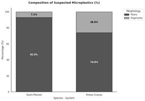
Figure 1. Distribution of
suspected microplastic morphology in fecal samples from goats and sheep.
The predominance of
fibers is consistent with reports that fibrous particles are frequent in
agricultural and livestock environments where textile residues, ropes, sacks,
plastic agricultural materials and other synthetic fibers may be present
(Mohammadi et al., 2025; Sheriff et al., 2023; Weithmann et al., 2018).
Nevertheless, the present study did not analyze environmental sources or
polymer composition; therefore, the association between fibers and specific
contamination sources remains a plausible interpretation rather than a
demonstrated causal pathway.
The literature indicates
that manure, digestates and livestock compost can contribute to the
redistribution of microplastics into agricultural soils (Quilliam et al., 2023;
Rana et al., 2023). In the evaluated systems, the presence of suspected
particles in feces suggests that fecal material could participate in local
redistribution if used as organic amendment. This interpretation should be
tested through parallel analysis of manure, soil, pasture and water under the
same production conditions.
Climate and management
may influence the mobility of plastic particles in pastoral landscapes. Dry
environments can favor surface accumulation and wind resuspension of particles
(Evangelou et al., 2024; Rezaei et al., 2022), whereas humid environments can
facilitate transport by rainfall and runoff toward rivers, drainage channels
and flooded areas (Severe et al., 2025). These mechanisms help contextualize
the Manabí and Guayas systems, but they remain explanatory hypotheses because
the study did not directly measure microplastics in air, soil, runoff or water
bodies.
Particle color was
retained as a descriptive observation but was not presented as a separate
quantitative table because the available aggregated color values were not
consistent with the overall concentration of 4.95 MP/g. Without the original
particle-level database by color and species/system, it is not possible to
calculate total particles, percentages, mean +/- SD, exact p-values or effect
sizes for color categories. Accordingly, chromatic findings should be
interpreted only as visual tendencies: darker particles were more frequently
described in goat samples, whereas lighter particles were more frequently
described in sheep samples. Color alone does not identify source, polymer type
or exposure route.
Overall, the observed
morphology and color patterns generate hypotheses about exposure pathways;
confirmation requires simultaneous analysis of soil, water, forage and polymer
composition. The findings support the need for integrated monitoring of fecal
and environmental matrices in small ruminant systems on the Ecuadorian coast,
while avoiding causal claims about specific sources or sanitary implications
that were not directly evaluated in the present study.
Conclusions
This study
detected suspected microplastic particles in fecal samples from goats and sheep
in two coastal Ecuadorian systems. Similar mean concentrations were observed
between goats and sheep; however, species and site effects cannot be separated
in the current design because each species was associated with a different
province and production system.
A
predominance of fibers over fragments was identified among the recovered
particles. Nevertheless, this finding should be interpreted as a morphological
pattern of the suspected particles observed in feces rather than as direct
evidence of specific contamination sources, since environmental matrices such
as soil, water, forage and feed were not analyzed simultaneously.
Chromatic
differences may reflect different environmental sources, but this should be
tested with environmental sampling and polymer identification. Therefore, the
observed color patterns should be considered exploratory and useful for
generating hypotheses about possible exposure pathways, rather than confirming
terrestrial, aquatic or atmospheric sources.
The
flotation/digestion protocol allowed preliminary isolation and morphological
characterization of suspected microplastic particles in fecal samples; however,
its analytical performance should be validated in future studies. Polymer
confirmation using FTIR or Raman spectroscopy is recommended to strengthen
particle identification and reduce uncertainty associated with visual
classification by light microscopy.
The findings
support the potential use of small ruminants as sentinels of environmental
exposure; future research should evaluate toxicological effects and possible
transfer to animal-derived products. Further studies should also incorporate
environmental matrices, standardized digestion procedures and confirmatory
polymer analysis to better understand the origin, distribution and implications
of suspected microplastics in pastoral livestock systems.
Conflict
of interest
The authors
declare that there is no conflict of interest.
Contribución
de los autores
Conceptualization: I.G.P., G.G.E., D.P.C.;
Methodology: I.G.P., E.V.M., B.A.P.; Investigation: I.G.P., E.V.M., B.A.P.;
Formal Analysis: I.G.P., G.G.E., D.P.C.; Writing—Original Draft Preparation:
I.G.P., G.G.E., E.V.M., B.A.P., D.P.C.; Writing—Review and Editing: I.G.P.,
B.A.P, G.G.E.; Supervision: D.P.C., I.G.P.
Referencias
bibliográficas
1.
Aransiola, S.
A., Babaniyi, B. R., Maddela, N. R., & Bello, O. S. (2025). White pollution:
Biodiversity and hazards in marine plastisphere. In White pollution:
Biodiversity and hazards in marine plastisphere (pp. 209–235). Springer Nature.
https://doi.org/10.1007/978-3-031-95547-1_10
2. Beriot, N., Peek, J., Zornoza, R.,
Geissen, V., & Huerta Lwanga, E. (2021). Low density microplastics detected
in sheep faeces and soil: A case study from intensive vegetable farming in
Southeast Spain. Science of the Total Environment, 755, 142653. https://doi.org/10.1016/j.scitotenv.2020.14265
3. Caicedo Olvera, J. A., & Paguay
Paguay, E. G. (2024). Evaluación de la presencia de microplásticos en fuentes
de agua y tipos de dieta en ovinos en Guayas, Ecuador [Trabajo de titulación
previo a la obtención del título de Médico Veterinario, Universidad de
Guayaquil]. Repositorio Institucional UG. https://repositorio.ug.edu.ec/handle/redug/7505
4. Celi-Simbaña, S. S., Andrade-Mora,
D. S., Loza-Pavón, S. J., & Bermeo-Sierra, T. I. (2023). Microplásticos, un
problema de salud pública emergente. Revista Información Científica, 102. https://doi.org/10.5281/zenodo.8105111
5. Evangelou, I., Tatsii, D.,
Bucci, S., & Stohl, A. (2024). Atmospheric resuspension of microplastics from bare
soil regions. Environmental Science & Technology, 58(22), 9741–9749.
https://doi.org/10.1021/acs.est.4c01252
6. González-Puetate, I. R., Martínez
Cepeda, G. E., Torres Lasso, P., Chávez Toledo, K. N., & Guevara Enrique,
G. (2024). Microplásticos en materia fecal de rumiantes en Ecuador. Ciencia
Veterinaria, 26(2), 114–129. https://doi.org/10.19137/cienvet202426203
7. Kanwal, H., Nadeem, H.,
Azeem, F., Rasul, I., Muzammil, S., Zubair, M., Imran, M., Afzal, M., &
Siddique, M. H. (2023). Generation and impact of microplastics and nanoplastics
from bioplastic sources. In Biodegradability of conventional plastics (pp.
83–99). Elsevier. https://doi.org/10.1016/B978-0-323-89858-4.00003-8
8. Landrigan, P. J., Raps,
H., Cropper, M., Bald, C., Brunner, M., Canonizado, E. M., … Dunlop, S. (2023).
The Minderoo Monaco Commission on plastics and human health. Annals of Global
Health, 89(1). https://doi.org/10.5334/aogh.4056
9. Mohammadi, M., Dargahi,
A., Almasi, A., Mousavi, S. A., & Mohammadi, P. (2025). Occurrence of
microplastic, antibiotics, hormones and heavy metals in livestock and poultry
manure in west Iran. Scientific Reports, 15(1), 27228.
https://doi.org/10.1038/s41598-025-03826-7
10.
Quilliam,
R. S., Pow, C. J., Shilla, D. J., Mwesiga, J. J., Shilla, D. A., &
Woodford, L. (2023). Microplastics in agriculture: A potential novel mechanism
for the delivery of human pathogens onto crops. Frontiers in Plant Science, 14.
https://doi.org/10.3389/fpls.2023.1152419
11.
Rana, M. M.,
Haque, M. R., Tasnim, S. S., & Rahman, M. M. (2023). The potential
contribution of microplastic pollution by organic fertilizers in agricultural
soils of Bangladesh: Quantification, characterization and risk appraisals.
Frontiers in Environmental Science, 11.
https://doi.org/10.3389/fenvs.2023.1205603
12.
Rezaei,
M., Abbasi, S., Pourmahmood, H., Oleszczuk, P., Ritsema, C., & Turner, A.
(2022). Microplastics in agricultural soils from a semi arid region and their
transport by wind erosion. Environmental Research, 212, 113213.
https://doi.org/10.1016/j.envres.2022.113213
13.
Severe,
E., Surridge, B. W. J., Fiener, P., Coogan, M. P., Platel, R. H., James, M. R.,
& Quinton, J. (2025). The transport of microplastics from soil in response
to surface runoff and splash erosion. Environmental Science & Technology,
59(27), 14063–14074. https://doi.org/10.1021/acs.est.5c04795
14.
Sheriff,
I., Yusoff, M. S., Manan, T. S. B. A., & Koroma, M. (2023). Microplastics
in manure: Sources, analytical methods, toxicodynamic and toxicokinetic
endpoints in livestock and poultry. Environmental Advances, 12, 100372.
https://doi.org/10.1016/j.envadv.2023.100372
15.
Urli,
S., Corte Pause, F., Crociati, M., Baufeld, A., Monaci, M., & Stradaioli,
G. (2023). Impact
of microplastics and nanoplastics on livestock health: An emerging risk for
reproductive efficiency. Animals,
13(7), 1132. https://doi.org/10.3390/ani13071132
16.
Weithmann,
N., Möller, J. N., Löder, M. G. J., Piehl, S., Laforsch, C., & Freitag, R.
(2018). Organic fertilizer as a vehicle for the entry of microplastic into the
environment. Science
Advances, 4(4). https://doi.org/10.1126/sciadv.aap8060